A group of researchers has managed to create the most detailed and complete map of the human brain to date.
BigBrain, the 3D digital reconstruction of the brain of a 65-year-old woman, reveals its details with microscopic precision.
The brain is made up of numerous networks of neurons that vary enormously in size, shape, and layers. Scientists can now see cerebral neuronal distribution with greater detail using this new model.
The finding, published in the journal Science, could drive further research into a wide range of brain diseases, including Alzheimer’s and Parkinson’s.
BigBrain will serve as an atlas for neurosurgery and give researchers invaluable insight into how the brain processes emotion, cognition and language.
Lead author, Katrin Amunts, from the Research Centre Jülich, Germany, said that “it is a common basis for scientific discussions because everybody can work with this brain model.”
The brain model “redefines traditional maps from the beginning of the 20th century,” says Katrin.
Senior editor of the journal Science, Peter Stern, said that the scientists have pushed the limits of current technology.
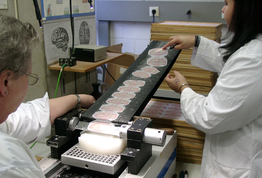
With a microtome, scientists cut the woman’s brain into tiny 20 micrometer-thick sections.
Courtesy: AMUNTS, ZILLES, EVANS ET AL.
In order to create the brain map, the scientists used 7,400 of the brain slices and assembled images of all of them to create a coherent three-dimensional volume of the brain. They used a high-resolution flatbed scanner to digitize the sections into a fully reconstructed high-resolution 3-D brain model.
According to Alan Evans, a professor at the Montreal Neurological Institute at McGill University in Montreal, Canada, the project was “a tour-de-force to assemble images of over 7400 individual histological sections, each with its own distortions, rips and tears, into a coherent 3-D volume.”
The BigBrain provides an unparalleled resolution of brain components. Stern said that the “the spatial resolution the researchers achieved exceeds that of presently available reference brains by a factor of 50.”
Amunts added that of course the team “would love to have spatial resolution going down to 1 micrometer,” but currently “there are simply no computers at this moment which would be capable to process such data, to visualize this or to analyze it.”
The team said that it has been “a dream for almost 20 years. The dream came true because of an interdisciplinary and intercontinental collaboration spanning from Europe to Canada and from neuroanatomy to supercomputing.”
Even though the atlas map was created using only one person’s brain it still provides important information to help interpret other brains, said David Van Essen, a neurobiologist at Washington University in St Louis, Missouri. “Getting a really accurate map in one individual is, I think, very valuable,” he added.
The researchers hope to use this map to extract measurements of cortical thickness and further understand how neurodegenerative disorders develop.
One day Amunts and her team hope to develop a brain model at a resolution capable of capturing every single detail of cell morphology – which would require a resolution of 1 micron.
Last year, a team of British and American scientists created a complete statistical picture of the human brain’s complex network. The simple mathematical model not only helped further understand healthy brains, but it also offered unique insights into schizophrenia and other disorders.
Written by Joseph Nordqvist