
Magnetic resonance imaging (MRI) is a noninvasive type of imaging test that healthcare professionals use to detect multiple sclerosis (MS) activity in the brain and spinal cord.
MRI scans use strong magnetic fields, radio waves, and computers to create detailed images of the body’s organs and soft tissues.
An MRI scan can detect MS activity
This article will explain how MS appears on an MRI scan and how often a person with MS should undergo MRI scans.
MS activity appears on an MRI scan as either bright or dark spots. Typical MS lesions tend to be oval or frame shaped. MS lesions can appear in both the brain’s white and gray matter.
Healthcare professionals may use a chemical contrast dye called gadolinium to
In MS, the immune system attacks and damages the protective myelin coating that surrounds the nerves. Healthcare professionals refer to this damage as lesions.
MRI scans can identify lesions that occur due to MS. MS lesions can show white matter inflammation, demyelination, and scarring, or sclerosis. Scans can let healthcare professionals know when lesions are new and growing and potentially how damaging they are to the brain.
Over 90% of people with an MS diagnosis have it confirmed by an MRI scan.
An MRI scan can reveal several things about a person’s MS, including:
- if there is a relapse in MS
- if the MS is active without worsening
- if the MS is stable without activity
- if there is new active inflammation
- if there is no evidence of activity
The results of an MRI scan will look different depending on the type of MS that a person has.
Clinically isolated syndrome
A person with clinically isolated syndrome (CIS) is experiencing the first episode of symptoms that occur due to inflammation and demyelination in the central nervous system. The symptoms of CIS will last for at least 24 hours.
CIS may or may not cause lesions that appear on an MRI scan.
CIS does not always progress to another form of MS. However, people with MS-like brain lesions that appear on an MRI scan have a 60–80% chance of going on to develop another form of MS.
Relapsing-remitting MS
With relapsing-remitting MS (RRMS), an MRI scan will show at least two separate areas of damage that have occurred at different points in time.
Primary progressive MS
People with primary progressive MS (PPMS) tend to have fewer brain lesions, and the lesions tend to contain fewer inflammatory cells. They also tend to have more lesions in the spinal cord than people with other forms of MS.
A
Secondary progressive MS
Secondary progressive MS (SPMS) is a form of MS that can occur in people who have had RRMS, and it features a general worsening of symptoms over time.
While a person’s symptoms become more severe, MRI scans will not tend to show an increase or growth in inflammation. The worsening of symptoms is due to the nerve damage that has already occurred.
An MRI scan is a noninvasive imaging test that healthcare professionals use to produce images of the body’s soft tissue and organs. MRI scans do not use radiation.
The technician obtains the scan using a large, tube-shaped magnet. The powerful magnet combines with computer-generated radio waves to create detailed images of the body’s soft tissue and organs.
There are no known risks associated with exposure to these types of strong magnetic fields.
Various types of MRI scans can monitor MS activity in the brain. Healthcare professionals can carry out different types of scans during the same MRI session.
T-1 weighted scan with gadolinium
T-1 scans can involve the use of gadolinium, which is a contrast dye, to look for new or growing lesions.
Permanently damaged areas of the brain appear as dark spots. These are also known as black holes or hypointense lesions.
Areas of new active inflammation in the brain appear white on T-1 scans.
New or expanding lesions captured by a T-1 scan might indicate that a person’s MS is worsening. According to
T-1 weighted scan without gadolinium
A T-1 weighted scan without contrast dye can show hypointense lesions, which may indicate areas of permanent nerve damage.
T-2 weighted scan
T-2 scans show the total number of old and new lesions in the brain from the onset of MS.
New MS lesions appear as bright spots on a T-2 scan. These are also known as hyperintense lesions.
Fluid-attenuated inversion recovery
Fluid-attenuated inversion recovery imaging reduces interference from the spinal fluid to help view the effects of MS. Anomalies remain bright, while normal brain fluid looks dark.
Spinal cord imaging
Spinal cord imaging identifies MS lesions in the spinal cord.
Spinal cord imaging can show that damage has occurred in different parts of the central nervous system at different points in time.
MRI scans cause a small amount of the body’s water protons to line up with their powerful magnetic field. Radio waves then pulse through the body, causing the protons to spin out of order.
Once the technician turns the radio waves off, the protons fall back to their original order. As they return to their original positions, the protons release signals that transmit to a computer. The computer then converts these signals to detailed 2D and 3D images of body tissue and organs.
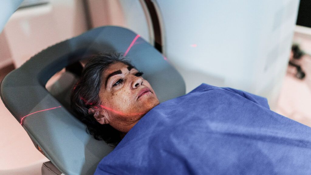
The MRI machine resembles a large tube with an examination table in the middle.
Before undergoing an MRI scan, a person needs to remove any clothing or personal items that may contain metal.
A healthcare professional places a padded covering partially over the person’s head to help keep it from moving during the scan.
The MRI machine makes loud knocking noises during the test. A person undergoing a scan will receive earplugs or earphones to help muffle the noise.
Gadolinium is a substance that forms the base of contrast dyes. It can only enter the brain if there is active inflammation. Inflammation from a new MS brain lesion breaks down the blood-brain barrier, allowing the gadolinium to leak into the brain.
Any gadolinium deposits that healthcare professionals find on an MRI scan suggest that there is disease activity in the brain.
The body almost completely clears gadolinium from the central nervous system after 48 hours.
Healthcare professionals use MRI scans to confirm a suspected diagnosis of MS. The first MRI scan helps serve as a comparison scan, especially in evaluating CIS.
A person with MS may expect to have routine monitoring of their condition every
- how long the person has had MS for
- any other medical conditions they have
- what their current treatment plan is
MRI scans use strong magnetic fields and radio waves to create detailed images of the central nervous system in individuals with MS. It is a safe and noninvasive test.
Healthcare professionals typically use MRI scans to both diagnose MS and to help monitor how a person responds to treatment.
For resources, research, and news for people living with MS, visit our dedicated MS hub.