
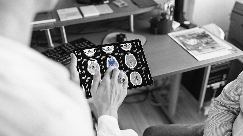
Doctors may use an MRI scan to detect a brain aneurysm in specific cases. MRI scans and other imaging techniques can help detect a brain aneurysm and reveal its shape and precise location.
A brain aneurysm is a weak area in a brain artery that swells and fills with blood. If the artery ruptures, it may cause severe brain injury or stroke and can be life threatening.
An MRI is one of the diagnostic tools doctors may use to detect some brain aneurysms. They may also use a CT scan or another type of imaging called a cerebral angiogram, which involves the injection of a contrast dye.
This article examines whether doctors use MRI scans to diagnose brain aneurysms and under what circumstances. It also discusses how accurate MRI scans are for brain aneurysms alongside the procedure and results of a brain MRI.
Finally, it looks at other tests to diagnose a brain aneurysm and answers some frequently asked questions.
An MRI machine uses a powerful magnetic force and a radio frequency to produce detailed images of soft tissues and structures in the body. To detect a brain aneurysm, an MRI may involve the injection of a contrast dye to highlight the arteries in the brain, which is a process called angiography.
Doctors will typically use an MRI angiogram to detect brain aneurysms that have not ruptured.
If doctors believe a brain aneurysm has ruptured and caused a bleed in the brain, known as a hemorrhage, they may opt for a CT angiography scan. Doctors may follow this with further tests.
MRI scans, especially with angiography, are generally
In a 2016 study, MRI scans detected
However, aneurysms of this size are less likely to rupture and lead to hemorrhage. The risk of hemorrhage is greater with aneurysms larger than 7 mm.
An MRI scan of the head can take 15–90 minutes, depending on how many images the MRI technicians are taking and the size of the area they are scanning.
Before an MRI scan, a person may receive an intravenous injection of contrast dye fluid.
They will then lie on the scanning table, and technicians will provide headphones or earplugs to block out the machine’s loud tapping noises. The scanning table is motorized and moves into the scanner, which is a short, open-ended cylinder.
Once a person is inside the MRI scanner, they and the MRI technician can communicate via an intercom system.
A person should remain still during the scan, and the MRI technician may instruct them to hold their breath at certain times.
The scanning process is painless. However, some people may experience anxiety and discomfort due to the loud noises and close confines of the MRI scanner. A doctor may provide a sedative to help a person relax during the procedure.
It may take a week or longer for a person to receive the results of an MRI.
If an MRI detects an unruptured brain aneurysm, a doctor will determine treatment options depending on a variety of factors. These
- the risk of the aneurysm rupturing
- the size, location, and type of aneurysm
- the risks involved in treatment
- the age, overall health, and medical history of the person
For small aneurysms that do not present a high risk of rupturing, doctors may monitor the condition with MRI or CT angiography scans.
The aneurysm may not require active treatment, and doctors may instead focus on treating any coexisting risk factors to reduce the risk of complications.
If an MRI scan detects higher risk or ruptured aneurysms, treatment may involve:
- Surgery: Doctors may perform an open brain surgery to stop blood flow to the aneurysm, called microvascular clipping. They may also implant a small stent to reroute blood flow or insert tiny wire coils to block blood flow to the aneurysm.
- Medication: Doctors may prescribe medication such as calcium-channel blockers to reduce the risk of stroke and anticonvulsive medication to prevent seizures from a ruptured aneurysm.
There are several tests doctors may use to diagnose a brain aneurysm in combination with or in place of an MRI scan.
These include:
- CT scan: Doctors may use a CT angiography scan if they suspect a brain aneurysm has ruptured. A CT scan provides two-dimensional images that a computer can combine into a more detailed, three-dimensional image.
- Cerebral angiography: A doctor will inject contrast dye through a catheter in the groin arteries. A person will then receive X-rays that outline the blood vessels so doctors can see weak spots in the arteries and identify aneurysms.
- Cerebrospinal fluid analysis: Doctors may perform a lumbar puncture, which people also call a spinal tap. They will use a thin needle to remove fluid from the lower spine for laboratory testing. This can help identify whether there is bleeding in the brain.
Below are the answers to some common questions about MRI scans and brain aneurysms.
Is a CT or MRI better to diagnose a brain aneurysm?
An MRI scan may be more effective in the diagnosis of unruptured brain aneurysms. However, a CT scan may be more effective in the diagnosis of ruptured aneurysms.
What is the survival rate of a brain aneurysm?
If a brain aneurysm does not rupture, a person can live a typical life.
The mortality rate for ruptured brain aneurysms is
An MRI scan is one diagnostic tool a doctor may use to diagnose a brain aneurysm. Doctors most commonly use MRI angiography scans, which include the use of a contrast dye, to diagnose unruptured brain aneurysms.
Doctors may use imaging techniques like CT angiography scans and cerebral angiography X-rays to diagnose higher-risk and ruptured aneurysms.
Depending on the results of an MRI scan, a person may require monitoring without active treatment or surgery and medication to treat a brain aneurysm.