
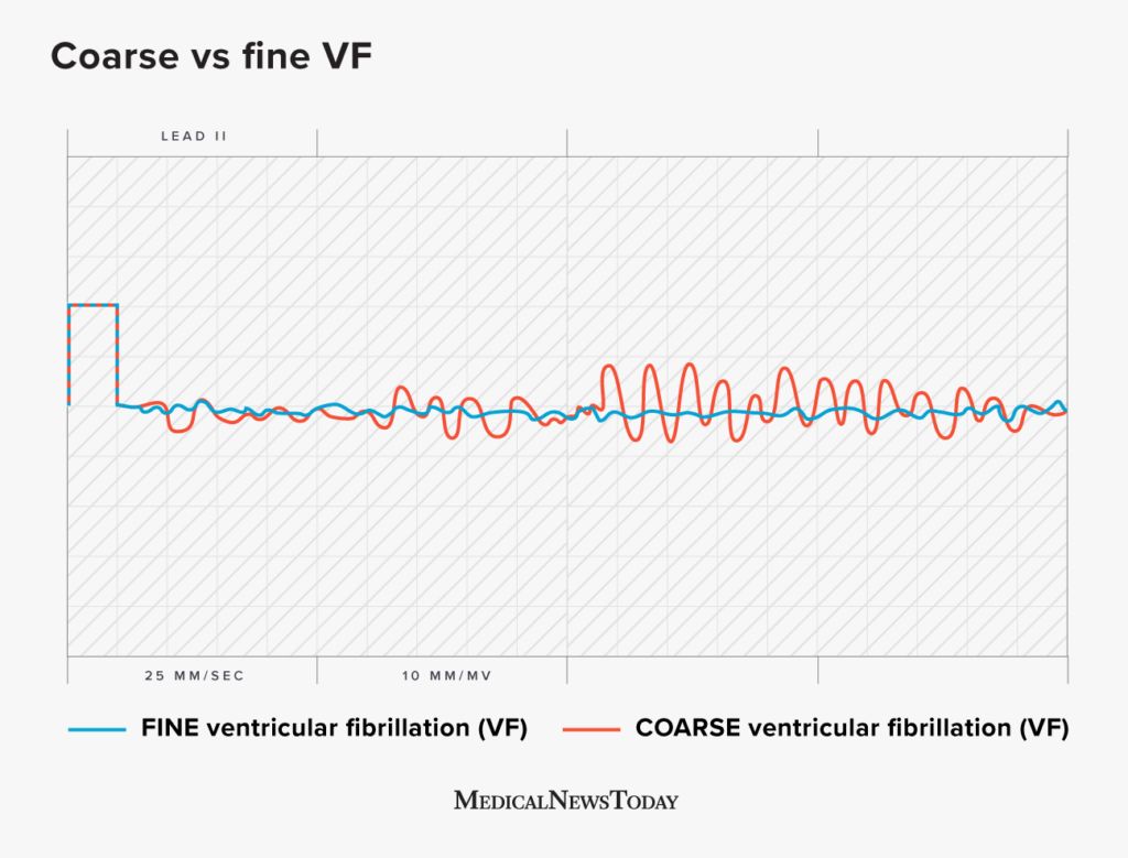
Depending on how the heart’s electrical activity appears on an EKG, a doctor may diagnose coarse or fine ventricular fibrillation (VF). Coarse VF appears as larger waves on an EKG compared with the waves relating to fine VF.
VF is an atypical heart rhythm that can be fatal without immediate treatment.
The ventricles are the lower chambers of the heart. Atypical electrical activity in the heart can cause the ventricles to quiver rather than beat naturally. This prevents the heart from pumping blood to the body, leading to cardiac arrest.
This article looks at the difference between coarse and fine VF and the symptoms, causes, diagnosis, treatment, and outlook.
Doctors use an EKG to show the heart’s electrical activity as it beats. The electrical signals appear as a wave on a graph and can show doctors if people have an irregular heartbeat.
As the heart beats, a wave passes through the heart, causing the heart to contract and pump blood. An EKG shows these waves, which indicate how fast the heart is beating. The waves appear as peaks and valleys.
A
Both fine and coarse VF are medical emergencies.
Fine VF may indicate a more negative outlook than coarse VF. This is because fine VF can indicate a worsened state. Coarse VF can also progress to fine VF as the condition worsens or if it continues for a prolonged period.
Prompt treatment is essential for both fine and coarse VF successful outcomes. Defibrillation may be more effective for coarse VF compared with fine VF.
Prompt defibrillation has a survival rate of
Signs of coarse VF and fine VF will appear on an EKG, which doctors identify
VF may cause the following symptoms:
- chest pain
- shortness of breath
- nausea and vomiting
- swelling in the lower legs
- sudden collapse
- loss of consciousness
- no pulse
Causes of VF
- lack of blood flow to the heart
- heart attack
- chest pain, or angina
- disease or damage to the heart muscle
- toxicity due to drugs or alcohol
- problems affecting the aorta, the main blood vessel to the heart
- sepsis
- heart surgery
- congenital heart disease
- injury to the heart area
- severe electric shock
- electrolyte imbalances
Doctors will immediately assess if a person is responsive and whether they have a pulse. They will need to diagnose and begin treatment for VF
Doctors use an EKG to diagnose VF and to identify if people have the coarse or fine type.
After treatment for VF, doctors will assess a person’s medical history and check for the underlying causes.
After diagnosing VF, a doctor will administer immediate shock delivery using a defibrillator. A defibrillator delivers an electric pulse, which restores a typical heartbeat.
If VF continues despite defibrillation, the person experiencing cardiac arrest will undergo CPR, and doctors will administer medications such as epinephrine and amiodarone. It is possible to perform multiple defibrillation attempts if the heart arrhythmia, such as VF, is shockable.
While resuscitating a person, doctors will also address any other issues, such as:
- treating a collapsed lung
- ensuring the airways remain open
- correcting any electrolyte imbalances
- administering fluids
- draining fluid buildup from around the heart
The following are answers to common questions about arrhythmias.
What are the four shockable rhythms?
Shockable rhythms are arrhythmias that respond to treatment with a defibrillator, which delivers an electric shock to the heart to restore a typical heart rhythm.
The shockable rhythms
How can a person tell the difference between AF and VF?
Atrial fibrillation (AF) and VF are both arrhythmias. AF is
AF does not always cause symptoms, but if it does, people may experience an irregular heartbeat, heart palpitations, lightheadedness, shortness of breath, fatigue, and chest pain.
VF begins in the ventricles, the heart’s lower chambers. VF is a
Healthcare professionals distinguish between AF and VF using an EKG.
What is the difference between fine VFib and asystole?
Asystole is a state in which there is
VF produces waves on an EKG, as there is still electrical activity. Prolonged VF may progress to asystole.
Ventricular fibrillation (VF) is a life threatening arrhythmia. Coarse VF and fine VF refer to the type of waves that appear on an EKG.
If VF continues, coarse VF may deteriorate to fine VF, which may have a more negative outcome. People will require immediate defibrillation for both types of VF. Prompt treatment may help improve survival rates.