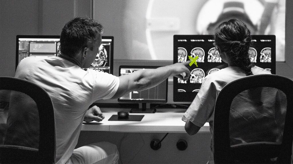
Doctors can use radiologic findings from MRI or CT scans to diagnose craniopharyngioma.
Craniopharyngioma is a benign, or noncancerous, tumor that grows on the pituitary gland. Often, people who develop this type of growth experience headaches, changes in vision, nausea, and vomiting.
A doctor may order an MRI or CT scan to diagnose craniopharyngioma. Once healthcare professionals identify the tumor, they can determine the next steps and treatment.
This article outlines how doctors use imaging to diagnose craniopharyngioma, treatment for craniopharyngioma, and when to speak with a doctor.

If a doctor suspects a person may have craniopharyngioma or another issue affecting the pituitary gland, they will likely refer them to radiology for image studies.
Radiology involves diagnosing and assessing various medical conditions using imaging, such as X-rays, CT, and MRI scans.
Doctors
MRI and CT scans are the two most common imaging tests healthcare professionals use to diagnose and assess craniopharyngioma.
MRI
An MRI uses strong magnetic fields to align protons in the body and create a detailed, three-dimensional image of its internal structures.
During a scan, a person lays as still as possible on a table that moves in and out of a large, enclosed tube. The test can take a few minutes to over an hour.
When diagnosing craniopharyngioma, the radiologist will likely inject a contrast dye. The dye helps provide a better image of the pituitary gland and potential growths or tumors. It can also help doctors view nearby structures, such as the hypothalamus or optic nerve.
A review of the surrounding structures can help doctors determine the tumor’s effects on the person’s health and guide treatment decisions.
An MRI does not use radiation and is generally safe for repeated imaging studies.
However, a person should inform the radiologist or technician if they have certain underlying health conditions. Someone with
- sensitive hearing, since an MRI can create loud noises
- implanted devices, particularly those with iron in them, such as pacemakers
- allergies to contrast dyes
- people with claustrophobia
- people who are pregnant
CT
A doctor may order or recommend a CT scan in addition to an MRI to help with diagnosis.
During a CT scan, a person will typically lie on a table. A machine will move a ring or circle around the head and neck area. Unlike an MRI, the CT scan does not completely enclose the person.
Craniopharyngioma tumors contain several components, including cystic properties, solid mass areas, and calcifications. A CT scan can show areas of calcification on the tumor.
The difference in calcification can help a doctor determine whether a person has the adamantinomatous or papillary subtype of this type of tumor.
Adamantinomatous subtype tumors are more common in children. Adamantinomatous craniopharyngioma tumors are usually large and irregular, with
Papillary craniopharyngioma tumors typically occur in adults and present as solid masses.
CT scans use X-rays to create an image. X-rays use ionizing radiation, which can potentially alter tissue. Though generally safe with occasional use, repeated CT scans
Children have an increased risk of cancer development from CT scans. Parents or caregivers may want to discuss the risk of CT scans with a doctor or radiologist.
Pregnant people should also discuss the risks of radiation exposure on the fetus with a healthcare professional before undergoing a CT scan.
Following an MRI or CT scan, a radiologist will review the images and share the results with the doctor who ordered them.
This doctor will typically review what the results mean with the person and help determine the next steps, which may include more testing or treatment.
Additional testing
MRI
An MRI can help doctors identify a tumor located on the pituitary gland. This test returns a detailed three-dimensional image of the pituitary gland, the presence of any tumors, and the surrounding structure.
The presence of a tumor can indicate craniopharyngioma. Its location and effects on surrounding tissue
CT scan
A CT scan can show calcification of the tumor. The presence of calcification can help a doctor determine the type of craniopharyngioma present.
A small 2016 study showed that
Treatment of craniopharyngioma is
Treatment options include surgery, radiation, and specific types of therapies.
Surgery may involve preoperative and postoperative hormone care, such as glucocorticoid replacement therapy. The most common procedure is endoscopic endonasal transsphenoidal or transcranial surgery. This generally involves the surgeon removing the tumor and working around surrounding structures and tissue.
Radiation may help reduce the rate of recurrence of tumors.
Intracystic therapy helps with cystic tumors. It involves injecting toxic substances to help shrink cysts. However, there is little evidence that this type of therapy is effective.
Following treatment, the survival rate at 5 years is between 80% and 95%. However, a person may experience a reduced quality of life due to endocrine-related health concerns, such as high blood pressure and fatigue.
A person should consider consulting a doctor if they experience symptoms that could indicate craniopharyngioma.
Some symptoms of craniopharyngioma include:
- visual disturbances
- nausea
- vomiting
- headaches
- endocrine-related issues, such as:
- abdominal weight gain
- high blood pressure
- mood swings
- purple marks on the skin
It is important to note that craniopharyngioma is rare. It has an estimated incidence of
Several other conditions may account for a person’s symptoms.
Doctors use radiology for craniopharyngioma diagnosis. An MRI can provide a clear picture of the tumor and its effects on surrounding tissue. A CT scan can help determine the type of tumor present.
A doctor may order additional testing to help determine the tumor’s effects on the person’s health.
Doctors often recommend surgical removal of the tumor in combination with radiation to prevent recurrence and hormone therapy to help with symptoms.
