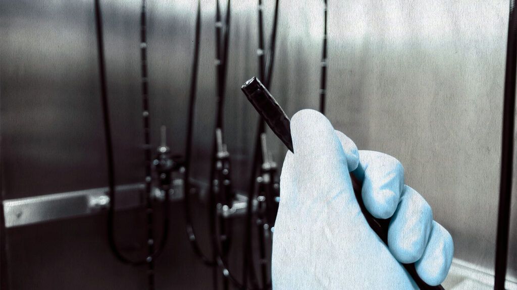
Esophagoscopy involves inserting a tube-like device — with a light and camera at its end — into the mouth and down the throat. The doctor can then examine the esophagus for abnormalities and take tissue samples (biopsy) if necessary.
The esophagus is the muscular tube that runs from the mouth to the stomach. Various disorders can affect it, including gastroesophageal reflux disease (GERD), cancer, strictures or narrowing, and esophagitis. An esophagoscopy is a medical procedure doctors use to explore and address these esophageal issues.
Sometimes, doctors can attach tools to the end of the device (known as an esophagoscope) to perform surgery or other interventions.
This article covers esophagoscopy, the various types, benefits, and what to expect during the procedure.

Esophagoscopy allows doctors to visually inspect the esophagus, obtain tissue samples for analysis, and treat specific issues.
It can help diagnose and/or treat the
The three types are:
Rigid esophagoscopy
This technique involves a rigid, straight instrument.
Doctors use rigid esophagoscopy when they need precise control during tissue sampling or minor surgeries, such as while treating
Flexible esophagoscopy
As the name suggests, this technique involves a flexible tool allowing greater maneuverability through the curved passages of the throat and esophagus.
Flexible esophagoscopy is particularly useful for the following:
- examining the upper portions of the throat and esophagus
- assessing conditions, such as GERD
- evaluating suspected structural abnormalities
Transnasal esophagoscopy
Doctors insert this tool through the nostril and down the back of the throat into the esophagus. It is a quick, minimally invasive procedure that does not usually require anesthetic.
Here is an overview of what typically occurs during the procedure:
- Anesthetic or sedation: Depending on the type of esophagoscopy, doctors administer anesthetic or sedation to ensure comfort during the procedure.
- Positioning: The person usually lies on their side. A mouthguard or bite block protects the teeth and the esophagoscopy instrument.
- Instrument insertion: The doctor gently inserts the esophagoscope through the mouth or nose and down the throat, guiding it into the esophagus. This may initially lead to some discomfort or a sensation of pressure.
- Visualization: As the esophagoscope advances, it provides direct visualization of the esophageal lining. The doctor carefully examines the esophagus and obtains tissue or performs surgery.
- Recovery: After the procedure, the team monitors the person while the effects of anesthetic or sedation wear off.
The healthcare team provides instructions, including:
- any dietary restrictions to adhere to
- when it is safe to resume eating and drinking
- when to continue medications
They also advise people on what to expect post-procedure, such as side effects to look out for and when to seek medical help.
Like any medical procedure, esophagoscopy carries the potential for risks and complications. These include:
- infection
- bleeding
- pain
- injury to the esophagus or nearby structures
- heart and lung problems
- sedation or anesthetic-related issues
- dental trauma
Minimizing complications during esophagoscopy is critical for the safety and well-being of the patient.
Doctors employ a range of strategies to do so, including:
- Careful patient assessment: This involves evaluation of the following:
- medical history
- current health status
- any underlying medical conditions or allergies
- current medications
- previous reactions to anesthetic or sedation
- Thorough preparation: The patient must fast to reduce the risk of aspiration, where the stomach contents enter the lungs. Proper hydration and electrolyte balance are also important.
- Precise technique: Experienced and skilled clinicians use well-maintained, sterile equipment to perform esophagoscopy. They also take precautions to minimize trauma to the esophageal lining during the insertion and removal of the esophagoscope.
- Continuous monitoring: Doctors closely monitor vital signs — including heart rate, blood pressure, and oxygen saturation — throughout the procedure.
- Post-procedure care: After esophagoscopy, the medical team monitors the individual for any signs of complications. They allow adequate time for recovery before discharging a patient.
Esophagoscopy offers many benefits:
- Noninvasive option: Esophagoscopy is relatively noninvasive compared with other surgeries. It often requires minimal incisions or anesthetic, resulting in a shorter recovery time and reduced discomfort.
- Supports precise diagnosis: Doctors can obtain tissue samples from the esophagus for detailed analysis and to facilitate diagnosis.
- Early detection and treatment: The procedure enables doctors to detect esophageal issues — such as cancer — earlier, allowing timely intervention and improved treatment outcomes. Sometimes, doctors can treat the issue at the same time, such as removing abnormal growths.
While esophagoscopy is generally safe for most people, there are some instances where doctors may not recommend it. These include when the person has:
- severe heart conditions
- severe respiratory distress
- had recent surgery on the esophagus or nearby structures
- had recent trauma to the neck or face
- active upper gastrointestinal bleeding
- any bleeding disorders
- esophageal perforation
- severe inflammation or swelling of the throat
Esophagoscopy allows doctors to diagnose and treat esophageal conditions. It involves inserting a tube with tools into the nose or mouth and into the esophagus, enabling visualization of the tissue. The tube may be rigid or flexible, depending on the procedure type.
It is generally a safe and effective procedure, with minimal recovery time and risk of complications.
