
A fiberoptic bronchoscopy is a medical procedure that allows a doctor to examine the airways, take samples for biopsy, remove objects from the airways, and perform minor treatments.
A fiberoptic bronchoscopy is an invasive medical procedure that a doctor may use to help diagnose respiratory health conditions, remove unwanted objects, and perform minor treatments, such as removing fluid from the airways.
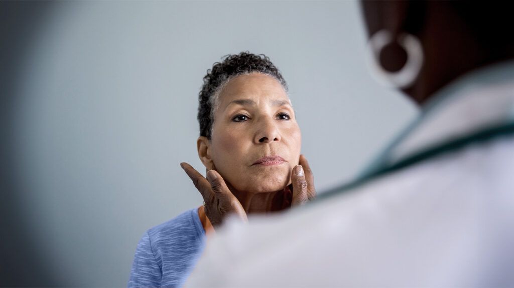
A doctor may recommend a flexible bronchoscopy if they believe a person has a respiratory condition, such as an infection, lung spots, or bleeding.
This article discusses fiberoptic bronchoscopy, when it might be useful, the technique a doctor may use, possible complications, and more.
A fiberoptic bronchoscope is a telescope with a long, flexible tube made of fiber optic material and a small light at the end. The fiber optic material consists of flexible glass fibers that can transmit light along the entire length of the telescope.
This can light up the inside of the airways and allow a doctor to see the lungs and airways clearly. The procedure is invasive, which means that it happens inside the body.
A doctor
Biopsy samples
A doctor may use fiberoptic bronchoscopy to take samples from inside the airways in order to perform various tests. Samples a doctor may take during a fiberoptic bronchoscopy can include:
- Lung cells: A doctor can pass a saltwater substance through the fiberoptic bronchoscope and into the lungs to collect cells. A doctor can then test them for the presence of infection or cancer cells.
- Lung tissue: A doctor may take biopsies of tissue from the lungs or airways by passing a small needle or forceps through the fiberoptic bronchoscope.
- Lymph nodes: A doctor may take samples of the lymph nodes in the lungs by passing a small needle or forceps through the fiberoptic bronchoscope.
A fiberoptic bronchoscopy can be useful when a doctor needs to see inside the airways to check for any irregularities or diagnose respiratory conditions. The doctor may want to take some samples of lung tissue or cells for further testing.
A doctor may also use fiberoptic bronchoscopy to treat an issue in the airways,
- removing an item that is stuck in the airways
- removing fluid from the airways
- cleaning out the airways
- widening an airway that has become narrow
- draining an abscess
At the start of a fiberoptic bronchoscopy, a doctor may administer sedation medication and local anesthetic.
Sedation medication can make a person feel relaxed and drowsy, which may make it easier to perform a fiberoptic bronchoscopy. Local anesthetic can numb the back of the throat.
A doctor can then pass the fiberoptic bronchoscope through the nose or throat, into the windpipe. The doctor may also pass a salt solution, tiny needles, brushes, or forceps through the fiberoptic bronchoscope to take biopsy samples.
Once the doctor completes the examination, they will slowly remove the fiberoptic bronchoscope.
A fiberoptic bronchoscopy may take approximately 20 minutes to complete. During the procedure, a person may feel some discomfort, a gagging sensation, or slight breathlessness as the tube passes through the windpipe.
Following fiberoptic bronchoscopy, a person may not be able to eat or drink for approximately 1 hour, until the anesthetic wears off. A person will typically go home the same day as the procedure.
A person may experience a sore throat or hoarse voice for several days or develop a fever for up to 24 hours following a fiberoptic bronchoscopy.
A person may also have a small amount of blood when they cough for 12–24 hours following the procedure. This typically occurs if a doctor takes tissue samples during the procedure.
Fiberoptic bronchoscopy is typically a safe procedure. Complications are usually minor — for example, having a sore throat for a few days afterward. However, it can sometimes lead to more serious complications,
- a collapsed lung due to the collection of lung tissue samples
- bleeding that lasts longer than 24 hours
- nerve damage, seizures, or a coma if there is an anesthetic overdose
- a chest infection, though this is rare
Here are some commonly asked questions about fiberoptic bronchoscopy.
Which is better: fiberoptic bronchoscopy or rigid bronchoscopy?
It depends on the health concern.
To diagnose conditions, take biopsy samples, and avoid having general anesthetic, a fiberoptic bronchoscopy is best. However, if a person has an object stuck in the airways, a rigid bronchoscopy has a
A rigid bronchoscopy can also open the airways more widely, allowing a person to breathe more easily. However, a person will need a general anesthetic so that they are asleep during the procedure.
What is a fiberoptic bronchoscope used for?
A fiberoptic bronchoscope
What is a fiberoptic bronchoscopy?
Fiberoptic bronchoscopy is a medical procedure
What are the advantages of fiberoptic bronchoscopy?
Fiberoptic bronchoscopy has several advantages. One advantage is that a person does not require general anesthesia and can stay awake during the procedure.
The procedure is typically safe and only takes approximately 20 minutes. A doctor can examine the airways visually as well as take biopsies, clear the airways, and perform minor treatments all in the same procedure.
Fiberoptic bronchoscopy is a medical procedure involving a long telescope that a doctor can use to examine the airways and diagnose any respiratory conditions.
This telescope consists of fiber optic materials, which light up the entire telescope. This allows a doctor to see clearly inside the airways. During the procedure, a doctor may take biopsy samples for further testing.
A doctor may use a fiberoptic bronchoscopy to remove an object that has become stuck in the airways, or for minor treatments such as removing fluid from the airways.
A person may experience complications. However, they are typically minor, such as having a sore throat for several days. More serious complications can involve bleeding and a collapsed lung.