
Achilles tendonitis (AT) is inflammation, pain, and swelling of the Achilles tendon. Insertional Achilles tendonitis is one type, which involves the lower part of the tendon that slots into the heel bone.
The Achilles tendon is a large tendon that runs down the back of the lower leg. It connects the calf muscles to the heel bone and helps with walking, running, and jumping.
With overuse, a person may experience tendonitis, also known as tendinitis. Insertional AT is one type of AT. It involves the lower portion of the tendon, where it connects to the heel bone.
This article explains insertional Achilles tendonitis, one of two types of AT inflammation.
Tendons connect the muscles to bones and joints,
Insertional AT is one of two types of Achilles tendon inflammation. This type involves the lower part of the tendon, where it inserts into the heel bone, also known as the calcaneus.
Inflammation is how the body responds to physical stress or injury. AT is inflammation of the Achilles tendon, and there usually is no link between it and a specific injury. Instead, AT develops due to repeated stress on the tendon.
Insertional AT can develop in anyone at any age, regardless of how much physical activity they do.
There are two types of Achilles tendinopathy: insertional and noninsertional.
Insertional AT pain occurs when the tendon attaches to the back of the heel bone. Noninsertional AT pain occurs slightly higher, roughly 2–6 centimeters (cm) above where the tendon attaches to the back of the heel bone.
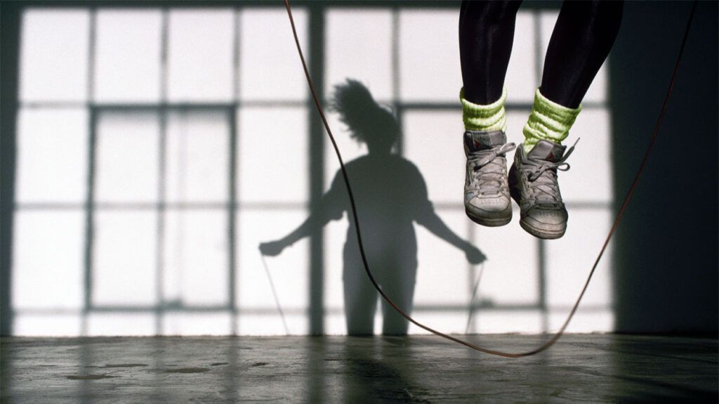
Noninsertional AT is the more common type and often occurs in younger people who engage in a lot of physical activity. Running is a significant contributing factor to both types of Achilles tendonitis.
A 2023 study of 3,379 runners found that
An individual can have both types at the same time.
Insertional AT does not typically relate to a specific injury. However, it is often the result of repetitive stress on the tendon.
Calf muscle tightness often causes insertional AT by increasing the stress on the Achilles tendon where it meets the heel. However, more general causes of AT can also lead to the insertional type.
For example, rapidly increasing exercise levels by extending the duration and intensity of workouts without giving the body time to adjust can increase inflammation.
Haglund’s deformity is another potential cause. This condition enlarges the bone at the back of the heel. This can, in turn, rub against the Achilles tendon, contributing to inflammation and pain.
Insertional AT symptoms
Symptoms include:
- pain and stiffness around the Achilles tendon area, especially upon waking
- Achilles tendon pain and heel pain that get worse during exercise
- increased pain the day after physical activity
- thicker tendons
- bone spurs
- swelling that gets worse during the day or with exercise
People with this type of AT might also struggle to put on footwear due to pain or discomfort at the back of the heel. Pain can last for longer than 3 months.
Once a person with suspected insertional AT visits a doctor with symptoms, the physician may look for the following physical signs:
- swelling of the Achilles tendon
- swelling at the back of the heel
- pain or bone spurs at the bottom of the tendon, which would be specific to insertional AT
- heel pain while stretching the calf
- ruling out pain in the middle of the tendon, which would point to noninsertional AT
- a reduced ability to point the toes downward
A doctor might also request one of several tests:
- X-rays, to have a closer look at the tendon
- MRI scans, if surgery is necessary to repair the tendon
- ultrasound, for some people, although it may produce less consistent results than an MRI, depending on the operator
At first, a doctor will recommend nonsurgical treatments.
Nonsurgical
Several nonsurgical approaches can help people with insertional AT manage tendon pain, including:
- Rest: It is vital to reduce or completely stop the activities that increase pain. This might involve switching from high impact exercises, such as running, to lower impact activities, including cycling and swimming.
- Ice: Apply an ice pack on the most painful region of the tendon for up to 20 minutes. This may help reduce pain and swelling. Try this as many times as necessary in a day.
- Nonsteroidal anti-inflammatory drugs (NSAIDs): Medications, including naproxen and ibuprofen, can provide relief from AT pain and swelling. These may allow increased movement for physical therapy exercises, even though they do not treat the underlying problem.
- Physical therapy: People with AT can perform various stretches and exercises under physical therapist supervision before continuing at home. These may cause discomfort at first.
- Wearing a splint while sleeping: This can help reduce pain upon waking for people with AT. During splinting, a removable brace holds the foot upward during sleep.
- Shoewear adjustments: Specialist shoes and orthotic insoles may help relieve pressure on the heel while walking.
- Extracorporeal shockwave therapy (ESWT): This therapy may speed up healing of the Achilles tissue using shockwaves, although more evidence is necessary to confirm its effectiveness as a routine AT treatment.
Surgical
Several types of surgery
These aim to remove damaged tissue or make the calf muscles longer. This can help reduce pain and improve function. Procedures might include:
- Debridement: The surgeon removes damaged tissue from the tendon. They will then reattach the remaining tendon to the heel bone using metal or plastic anchors. The timeline varies depending on the level of tendon damage, but most people can walk in a cast or surgical boot within 2 weeks.
- Gastrocnemius recession: The surgeon improves the motion of the ankle by lengthening the calf muscle. They can perform this by either open surgery or by making a smaller cut and inserting a tube with a camera attached.
- Minimally invasive surgery: The surgeon makes a small incision and removes damaged tissue using a thin, flexible camera and small surgical instrumentals. They may make small holes to release the tendon from the bone. More research is necessary to confirm the effectiveness of this procedure, though, especially for people with more severe tendonitis.
Achilles tendon surgery has enabled a return to pretendonitis function in around 75% of people who undergo the procedure, according to the American Association of Orthopaedic Surgeons.
After surgery, people may need up to a year of physical therapy to recover fully. Around 20–30% of people may continue to feel pain after surgery.
Insertional Achilles tendonitis is a type of AT. It occurs as a result of sustained pressure on the point at which the Achilles tendon slots into the heel bone. This usually develops as a result of excessive physical activity, causing bone spurs, pain, swelling, and restricted motion.
A range of nonsurgical treatment options are available, including rest, ice therapy, physical therapy, orthotic shoe adjustments, and splinting during sleep. If pain continues, surgery to remove damaged tissue or extend the calf muscle is often successful.