
Intraretinal microvascular abnormalities (IRMAs) are abnormal branching, or dilation, of the blood vessels in the retina. It occurs as a complication of diabetic retinopathy.
Diabetic retinopathy describes a potential ocular complication of diabetes. It refers to when persistently high blood sugar levels damage the delicate blood vessels of the eye’s retina.
This can cause the tiny blood vessels in the retina to leak or bleed. As the condition progresses, it can lead to severe vision impairment and, sometimes, irreversible blindness.
In this article, we explore IRMAs in more detail, including how it is different from neovascularization and treatment.
Intraretinal microvascular abnormalities (IRMAs) refer to a potential complication of diabetic retinopathy. Damage may start to occur in the blood vessels of the eyes if a person experiences long periods of high blood sugar levels. This damage can lead to IRMAs, which is when the blood vessels of the retina become stretched out or may grow in areas where they should not be present.
The presence of IRMAs typically indicates the end stage of nonproliferative diabetic retinopathy (NPDR).
NPDR describes the earlier stages of the condition before it progresses into the final and most advanced stage, known as proliferative diabetic retinopathy (PDR). The characteristic feature of PDR is the growth of new abnormal blood vessels, known as neovascularization.
Evidence notes that IRMAs are a known
Read on to learn more about the differences between NPDR and PDR.
Another complication of diabetic retinopathy is neovascularization. It is similar to IRMAs in that they both involve abnormal blood vessel growth. However, some key differences exist:
- Appearance: Typically, IRMAs will appear more disorganized and with sharper outlines. They also do not cross over major retinal blood vessels. However, neovascularization has a rounded outline, with a fan or branch-like structure, and sometimes crosses over major blood vessels. IRMAs are also often slightly larger than neovascularization.
- Location: Whereas IRMAs occur on the intraretinal layers of the eyes, neovascularisation is more centered or focal in location, depending on how severe it is, and it grows on top of the retina.
- Leakage: IRMAs tend to have little or no leakage, whereas neovascularization may cause the vessels to leak.
IRMAs may also be a precursor to neovascularization. The presence of IRMA suggests that neovascularization will soon occur on the surface of the retina or optic disk.
Read on to learn more about the stages of diabetic retinopathy.
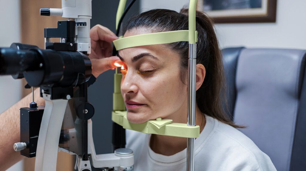
A healthcare professional, such as an ophthalmologist will initially do a dilated and comprehensive eye examination to assess for IRMA. This involves various tests to check vision, pressure, eye muscle function, and pupil response.
An eye doctor will also dilate a person’s pupils by using drops, so they can see any issues inside the eye. They may also use image tests, such as:
- a fundoscopy, which allows a doctor to view the back of the inside of the eye, including the retina
- optical coherence tomography (OCT), which uses light waves to take a cross-sectional picture of the retina to
help identify and monitor IRMAs - fluorescein angiography, which involves injecting a fluorescent dye into the bloodstream, allowing the ophthalmologist to use a camera to take pictures of the retina and assess blood flow.
An eye doctor may also use a combination tool, known as optical coherence tomography angiography (OCTA), to help distinguish between IRMAs and neovascularization.
Regular eye examinations are crucial for people living with diabetes to enable timely detection of IRMAs and other symptoms of diabetic retinopathy. Early diagnosis enables proactive management, preventing the progression of the condition and reducing the risk of vision loss.
Read on to learn more about diabetic eye screening.
Healthcare professionals may use the 4-2-1 rule to determine the severity of NPDR.
The 4-2-1 rule
- severe hemorrhages in all four quadrants of the retina
- venous beading in two or more quadrants of the retina
- moderate IRMAs in one or more quadrants of the retina
A person may have severe NPDR retinopathy if they display one of these symptoms and very severe NPDR if they fulfill two or more of these criteria.
Managing IRMAs and diabetic retinopathy may involve combining diabetes management and treatment to help stop the progression of retinopathy.
Managing diabetes largely focuses on managing blood glucose levels and
- taking medication as a doctor prescribes
- exercising regularly
- managing a person’s diet
- maintaining a suitable body weight
- checking blood glucose levels regularly
In terms of treatment for diabetic retinopathy and IRMAs, this will depend on the severity. Some available treatments
- Laser therapy: The procedure involves using a focused laser beam to seal the leaking blood vessels and prevent the growth of abnormal vessels.
- Injections: Antivascular endothelial growth factor (VEGF) medications can help reduce the growth of abnormal blood vessels and aid swelling within the retina. A doctor will administer this injection into the vitreous — the jelly-like substance in the eye.
- Eye surgery: In people with more advanced cases, where bleeding and scar formation are impacting vision, a doctor may recommend a procedure called a vitrectomy. During this procedure, a doctor will remove the vitreous and replace it with a clear fluid.
The impact of IRMAs on vision can vary depending on the stage of diabetic retinopathy. As IRMAs typically indicate severe NPDR or PDR, a person
- blurry vision
- floaters
- distorted vision
- partial vision loss
- total vision loss
Intraretinal microvascular abnormalities (IRMAs) are a complication of diabetic retinopathy. They occur due to damage to the delicate blood vessels in the retina.
IRMAs serve as a crucial indicator of abnormal blood vessel changes within the retina and suggest end stage nonproliferative diabetic retinopathy. IRMAs are also a risk factor for neovascularization, which is a characteristic feature of the most advanced stage of diabetic retinopathy, known as proliferative diabetic retinopathy.
Effective management of diabetic retinopathy and its complications involves a combination of regular monitoring, lifestyle modifications, and targeted treatments. Timely intervention is key to preserving vision and reducing the impact of diabetic retinopathy on the quality of life of individuals living with diabetes.