
Radiology tests can help doctors stage laryngeal cancer, determine treatment, and monitor progress.
Radiology uses imaging tests to help diagnose and treat diseases. They help stage laryngeal cancer and check if the cancer has spread.
This article examines radiology tests and how doctors use them for laryngeal cancer.
Doctors
- to examine areas that may be cancerous
- to identify if and how far the cancer has spread
- to check how well the treatment is working
- to check for any signs of the cancer returning after treatment
Doctors
- CT scan: A CT scan creates detailed images of the inside of the body with X-rays.
- MRI scan: An MRI scan uses radio waves and magnets to create detailed images of the body.
- PET scan: A PET scan uses fluorodeoxyglucose (FDG), a slightly radioactive substance. FDG collects in cancer cells and shows up on a PET scan.
- PET-CT scan: Doctors may use a PET-CT machine that combines both a PET and a CT scan. Using both these tests allows doctors to see areas of increased radioactivity due to the FDG alongside detailed CT images.
Other tests include:
- Chest X-ray: A chest X-ray checks if laryngeal cancer has spread to the lungs, although doctors usually use a CT scan for this as it provides more detail.
- Bone scan: A bone scan checks if laryngeal cancer has spread to the bones. A doctor injects a radioactive substance into the blood that collects in cancerous areas of bone. However, doctors tend to use a PET scan instead.
- Barium swallow: People swallow barium, a substance that lines the throat and esophagus and allows doctors to see clearer X-ray images. Doctors may do this if they have concerns that the gastrointestinal tract or a person’s breathing is affected.
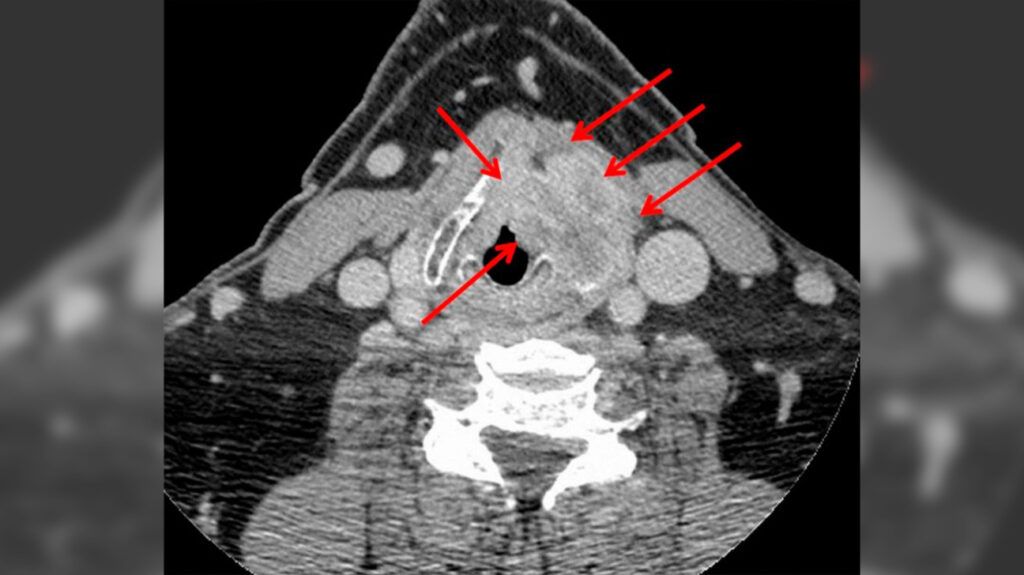
Doctors may use CT scans for early or late stage laryngeal cancer. They may use a contrast-enhanced CT scan that uses a contrast dye to create clearer images.
A CT scan can examine the tumor size and see if the cancer is spreading to the surrounding lymph nodes. It can also help stage laryngeal cancer by highlighting issues with the lymphatic system in the neck and other structures.
A
Additionally, a CT scan can provide more detailed results than a direct laryngoscopy. This is a procedure doctors use to examine the throat to diagnose laryngeal cancer.
If people receive a laryngeal cancer diagnosis, doctors
MRIs can help diagnose different tumors by providing detailed images of the soft tissues and showing the tumor stage and whether it affects the cartilage. This helps doctors decide on the best course of treatment for the specific tumor type.
Doctors may use gadolinium, a substance they inject into a vein, to provide stronger contrast and clearer images.
PET results can provide doctors with important information on the tumor and the surrounding area.
Doctors
- if they have found areas on other tests that may be cancerous
- to stage the cancer
- to check if the cancer has spread, in more advanced stages
- to help predict treatment responses and a person’s outlook
Doctors use PET to look at metabolic tumor volume, a feature that can help them predict progression-free survival. Progression-free survival refers to the length of time someone lives with the cancer without it worsening.
Doctors may use a PET-CT machine, which combines both a PET and CT scan. Using both these tests allows doctors to see areas of increased radioactivity due to the FDG alongside detailed CT images.
A doctor will provide instructions for preparing for radiology tests, which may include:
- avoiding food or drink for 2 hours or more before a scan
- removing any jewelry or metal objects
- letting a doctor know about any implants,
such as a pacemaker - informing a doctor of any potential allergies to contrast dyes
- taking a relaxant, if people have claustrophobia
The following are common questions about diagnosing laryngeal cancer.
What other tests do doctors use to diagnose laryngeal radiology?
Other diagnostic tests for laryngeal cancer
A panednoscopy examines the larynx, esophagus (food pipe), and windpipe. A biopsy removes a tissue sample to examine for cancer cells.
What is the most frequent site of carcinoma of the larynx and has the best outlook?
Around
Glottic cancer has the best outlook, with a
How curable is laryngeal cancer?
Stage 0 laryngeal cancer
For more advanced cancers, removing the larynx may treat the cancer, or treatment may help prevent the disease’s growth.
What are the first signs of laryngeal cancer?
Signs of laryngeal cancer include sore throat, coughing up blood, ear pain, difficulty swallowing, a lump in the neck, and voice changes.
Doctors may use radiology tests to help stage a laryngeal cancer diagnosis and check if the cancer has spread.
Tests include CT, MRI, and PET scans, which all help doctors see inside the body and view cancerous changes.