
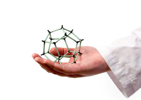
Nanotechnology, the manipulation of matter at the atomic and molecular scale to create materials with remarkably varied and new properties, is a rapidly expanding area of research with huge potential in many sectors, ranging from healthcare to construction and electronics. In medicine, it promises to revolutionize drug delivery, gene therapy, diagnostics, and many areas of research, development and clinical application.
This article does not attempt to cover the whole field, but offers, by means of some examples, a few insights into how nanotechnology has the potential to change medicine, both in the research lab and clinically, while touching on some of the challenges and concerns that it raises.
The prefix “nano” stems from the ancient Greek for “dwarf”. In science it means one billionth (10 to the minus 9) of something, thus a nanometer (nm) is is one billionth of a meter, or 0.000000001 meters. A nanometer is about three to five atoms wide, or some 40,000 times smaller than the thickness of human hair. A virus is typically 100 nm in size.
The ability to manipulate structures and properties at the nanoscale in medicine is like having a sub-microscopic lab bench on which you can handle cell components, viruses or pieces of DNA, using a range of tiny tools, robots and tubes.
Therapies that involve the manipulation of individual genes, or the molecular pathways that influence their expression, are increasingly being investigated as an option for treating diseases. One highly sought goal in this field is the ability to tailor treatments according to the genetic make-up of individual patients.
This creates a need for tools that help scientists experiment and develop such treatments.
Imagine, for example, being able to stretch out a section of DNA like a strand of spaghetti, so you can examine or operate on it, or building nanorobots that can “walk” and carry out repairs inside cell components. Nanotechnology is bringing that scientific dream closer to reality.
For instance, scientists at the Australian National University have managed to attach coated latex beads to the ends of modified DNA, and then using an “optical trap” comprising a focused beam of light to hold the beads in place, they have stretched out the DNA strand in order to study the interactions of specific binding proteins.
Meanwhile chemists at New York University (NYU) have created a nanoscale robot from DNA fragments that walks on two legs just 10 nm long. In a 2004 paper published in the journal Nano Letters, they describe how their “nanowalker”, with the help of psoralen molecules attached to the ends of its feet, takes its first baby steps: two forward and two back.
One of the researchers, Ned Seeman, said he envisages it will be possible to create a molecule-scale production line, where you move a molecule along till the right location is reached, and a nanobot does a bit chemisty on it, rather like “spot-welding” on a car assembly line. Seeman’s lab at NYU is also looking to use DNA nanotechnology to make a biochip computer, and to find out how biological molecules crystallize, an area that is currently fraught with challenges.
The work that Seeman and colleagues are doing is a good example of “biomimetics”, where with nanotechnology they can imitate some of the biological processes in nature, such as the behavior of DNA, to engineer new methods and perhaps even improve them.
DNA-based nanobots are also being created to target cancer cells. For instance, researchers at Harvard Medical School in the US reported recently in Science how they made an “origami nanorobot” out of DNA to transport a molecular payload. The barrel-shaped nanobot can carry molecules containing instructions that make cells behave in a particular way. In their study, the team successfully demonstrates how it delivered molecules that trigger cell suicide in leukemia and lymphoma cells.
Nanobots made from other materials are also in development. For instance, gold is the material scientists at Northwestern University use to make “nanostars”, simple, specialized, star-shaped nanoparticles that can href=”http://www.medicalnewstoday.com/articles/243856.php”>deliver drugs directly to the nuclei of cancer cells. In a recent paper in the journal ACS Nano, they describe how drug-loaded nanostars behave like tiny hitchhikers, that after being attracted to an over-expressed protein on the surface of human cervical and ovarian cancer cells, deposit their payload right into the nuclei of those cells.
The researchers found giving their nanobot the shape of a star helped to overcome one of the challenges of using nanoparticles to deliver drugs: how to release the drugs precisely. They say the shape helps to concentrate the light pulses used to release the drugs precisely at the points of the star.
Scientists are discovering that protein-based drugs are very useful because they can be programmed to deliver specific signals to cells. But the problem with conventional delivery of such drugs is that the body breaks most of them down before they reach their destination.
But what if it were possible to produce such drugs in situ, right at the target site? Well, in a recent issue of Nano Letters, researchers at Massachusetts Institute of Technology (MIT) in the US show how it may be possible to do just that. In their proof of principle study, they demonstrate the feasibility of self-assembling “nanofactories” that make protein compounds, on demand, at target sites. So far they have tested the idea in mice, by creating nanoparticles programmed to produce either green fluorescent protein (GFP) or luciferase exposed to UV light.
The MIT team came up with the idea while trying to find a way to attack metastatic tumors, those that grow from cancer cells that have migrated from the original site to other parts of the body. Over 90% of cancer deaths are due to metastatic cancer. They are now working on nanoparticles that can synthesize potential cancer drugs, and also on other ways to switch them on.
Nanofibers are fibers with diameters of less than 1,000 nm. Medical applications include special materials for wound dressings and surgical textiles, materials used in implants, tissue engineering and artificial organ components.
Nanofibers made of carbon also hold promise for medical imaging and precise scientific measurement tools. But there are huge challenges to overcome, one of the main ones being how to make them consistently of the correct size. Historically, this has been costly and time-consuming.
But last year, researchers from North Carolina State University, revealed how they had developed a new method for making carbon nanofibers of specific sizes. Writing in ACS Applied Materials & Interfaces in March 2011, they describe how they managed to grow carbon nanofibers uniform in diameter, by using nickel nanoparticles coated with a shell made of ligands, small organic molecules with functional parts that bond directly to metals.
Nickel nanoparticles are particularly interesting because at high temperatures they help grow carbon nanofibers. The researchers also found there was another benefit in using these nanoparticles, they could define where the nanofibers grew and by correct placement of the nanoparticles they could grow the nanofibers in a desired specific pattern: an important feature for useful nanoscale materials.
Lead is another substance that is finding use as a nanofiber, so much so that neurosurgeon-to-be Matthew MacEwan, who is studying at Washington University School of Medicine in St. Louis, started his own nanomedicine company aimed at revolutionizing the surgical mesh that is used in operating theatres worldwide.
The lead product is a synthetic polymer comprising individual strands of nanofibers, and was developed to repair brain and spinal cord injuries, but MacEwan thinks it could also be used to mend hernias, fistulas and other injuries.
Currently, the surgical meshes used to repair the protective membrane that covers the brain and spinal cord are made of thick and stiff material, which is difficult to work with. The lead nanofiber mesh is thinner, more flexible and more likely to integrate with the body’s own tissues, says MacEwan. Every thread of the nanofiber mesh is thousands of times smaller than the diameter of a single cell. The idea is to use the nanofiber material not only to make operations easier for surgeons to carry out, but also so there are fewer post-op complications for patients, because it breaks down naturally over time.
Researchers at the Polytechnic Institute of New York University (NYU-Poly) have recently demonstrated a new way to make nanofibers out of proteins. Writing recently in the journal Advanced Functional Materials, the researchers say they came across their finding almost by chance: they were studying certain cylinder-shaped proteins derived from cartilage, when they noticed that in high concentrations, some of the proteins spontaneously came together and self-assembled into nanofibers.
They carried out further experiments, such as adding metal-recognizing amino acids and different metals, and found they could control fiber formation, alter its shape, and how it bound to small molecules. For instance, adding nickel transformed the fibers into clumped mats, which could be used to trigger the release of an attached drug molecule.
The researchers hope this new method will greatly improve the delivery of drugs to treat cancer, heart disorders and Alzheimer’s disease. They can also see applications in regeneration of human tissue, bone and cartilage, and even as a way to develop tinier and more powerful microprocessors for use in computers and consumer electronics.
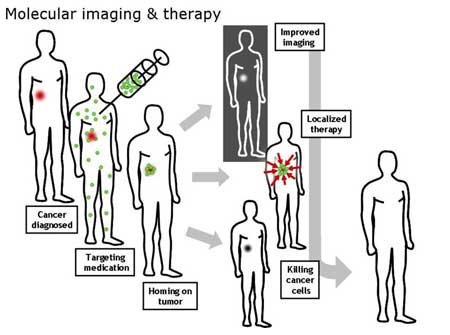
A schematic illustration showing how nanoparticles or other cancer drugs might be used to treat cancer. This illustration was made for the Opensource Handbook of Nanoscience and Nanotechnology
Recent years have seen an explosion in the number of studies showing the variety of medical applications of nanotechnology and nanomaterials. In this article we have glimpsed just a small cross-section of this vast field. However, across the range, there exist considerable challenges, the greatest of which appear to be how to scale up production of materials and tools, and how to bring down costs and timescales.
But another challenge is how to quickly secure public confidence that this rapidly expanding technology is safe. And so far, it is not clear whether that is being done.
There are those who suggest concerns about nanotechnology may be over-exaggerated. They point to the fact that just because a material is nanosized, it does not mean it is dangerous, indeed nanoparticles have been around since the Earth was born, occurring naturally in volcanic ash and sea-spray, for example. As byproducts of human activity, they have been present since the Stone Age, in smoke and soot.
Of attempts to investigate the safety of nanomaterials, the National Cancer Institute in the US says there are so many nanoparticles naturally present in the environment that they are “often at order-of-magnitude higher levels than the engineered particles being evaluated”. In many respects, they point out, “most engineered nanoparticles are far less toxic than household cleaning products, insecticides used on family pets, and over-the-counter dandruff remedies,” and that for instance, in their use as carriers of chemotherapeutics in cancer treatment, they are much less toxic than the drugs they carry.
It is perhaps more in the food sector that we have seen some of the greatest expansion of nanomaterials on a commercial level. Although the number of foods that contain nanomaterials is still small, it appears set to change over the next few years as the technology develops. Nanomaterials are already used to lower levels of fat and sugar without altering taste, or to improve packaging to keep food fresher for longer, or to tell consumers if the food is spoiled. They are also being used to increase the bioavailablity of nutrients (for instance in food supplements).
But, there are also concerned parties, who highlight that while the pace of research quickens, and the market for nanomaterials expands, it appears not enough is being done to discover their toxicological consequences.
This was the view of a science and technology committee of the House of Lords of the British Parliament, who in a recent report on nanotechnology and food, raise several concerns about nanomaterials and human health, particularly the risk posed by ingested nanomaterials.
For instance, one area that concerns the committee is the size and exceptional mobility of nanoparticles: they are small enough, if ingested, to penetrate cell membranes of the lining of the gut, with the potential to access the brain and other parts of the body, and even inside the nuclei of cells.
Another is the solubility and persistence of nanomaterials. What happens, for instance, to insoluble nanoparticles? If they can’t be broken down and digested or degraded, is there a danger they will accumulate and damage organs? Nanomaterials comprising inorganic metal oxides and metals are thought to be the ones most likely to pose a risk in this area.
Also, because of their high surface area to mass ratio, nanoparticles are highly reactive, and may for instance, trigger as yet unknown chemical reactions, or by bonding with toxins, allow them to enter cells that they would otherwise have no access to.
For instance, with their large surface area, reactivity and electrical charge, nanomaterials create the conditions for what is described as “particle aggregation” due to physical forces and “particle agglomoration” due to chemical forces, so that individual nanoparticles come together to form larger ones. This may lead not only to dramatically larger particles, for instance in the gut and inside cells, but could also result in disaggregation of clumps of nanoparticles, which could radically alter their physicochemical properties and chemical reactivity.
“Such reversible phenomena add to the difficulty in understanding the behaviour and toxicology of nanomaterials,” says the committee, whose overall conclusion is that neither Government nor the Research Councils are giving enough priority to researching the safety of nanotechnology, especially “considering the timescale within which products containing nanomaterials may be developed”.
They recommend much more research is needed to “ensure that regulatory agencies can effectively assess the safety of products before they are allowed onto the market”.
It would appear, therefore, whether actual or perceived, the potential risk that nanotechnology poses to human health must be investigated, and be seen to be investigated. Most nanomaterials, as the NCI suggests, will likely prove to be harmless.
But when a technology advances rapidly, knowledge and communication about its safety needs to keep pace in order for it to benefit, especially if it is also to secure public confidence. We only have to look at what happened, and to some extent is still happening, with genetically modified food to see how that can go badly wrong.
Written by Catharine Paddock PhD