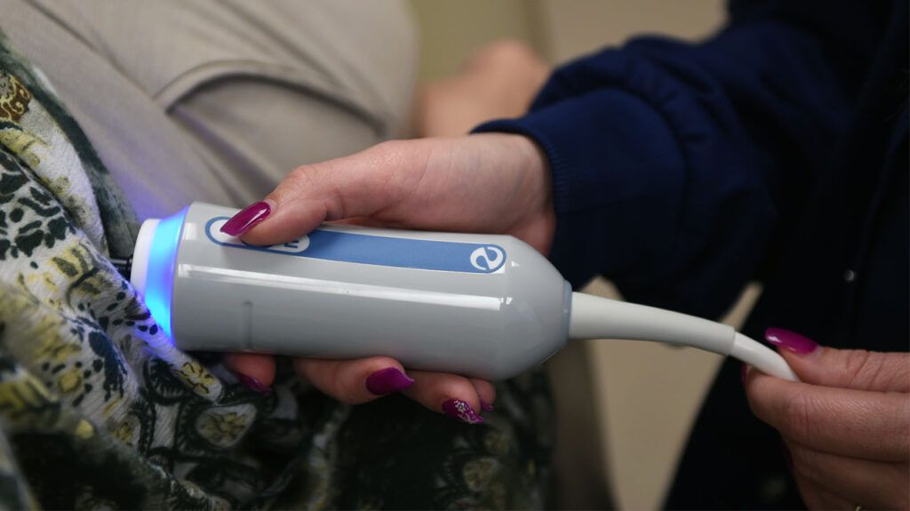
FibroScan is a diagnostic test that uses ultrasound to visualize the liver. Detecting cancer is not its main use. However, because the scan evaluates overall liver health, it may provide an indication.
FibroScan allows doctors to assess the liver without using invasive procedures. The test bounces sound waves off the liver tissue to gauge its characteristics.
The scan can show signs of liver damage, such as fat buildup or scarring. These findings can indicate a person is at a higher risk of liver cancer, but it does not confirm liver cancer on its own.
This article examines FibroScan as a cancer detection tool, how it works, and the procedure.

FibroScan is a test that can evaluate liver stiffness, fat buildup, and scarring. Doctors can also use it to monitor liver changes over time, which may indicate if a person is at risk of liver cancer.
For example, there is an
A 2020 study explored cancer diagnoses in people with cirrhosis, or advanced scarring, due to hepatitis C. The researchers noted that those who had liver stiffness of 24 kilopascals (kPa) or more in a FibroScan were more at risk of cancer.
It is also possible for liver tumors to affect the results of a FibroScan, causing a higher score. However, only a liver biopsy can confirm cancer.
FibroScan can detect cirrhosis and other forms of liver damage. It can also diagnose and track
FibroScans use elastography to evaluate liver health. A specialized probe sends low-frequency sound waves into the liver, and the device calculates the speed at which these waves travel through the tissue. The resulting measurement quantitatively assesses liver stiffness.
FibroScan cannot accurately confirm or rule out cancer, but its accuracy for assessing liver health more generally is well-documented in
The United Kingdom’s National Health Service (NHS) notes that in around 1 in 10 cases, accurate FibroScan results are difficult to obtain. Certain conditions can interfere with FibroScan results, such as:
- ascites, or fluid buildup in the abdomen
- scarring from previous surgeries or radiation treatment
- obstructions inside bile ducts
Liver inflammation or congestion may also produce a result that is too high. In cases where the result is unclear or unreliable, a doctor may recommend a liver biopsy.
Some evidence has suggested that FibroScan measurements can be different in those with a higher BMI. However, a
The FibroScan procedure is as follows:
- Preparation: Before the FibroScan procedure, a person should not eat or drink for 3 hours to ensure optimal conditions for accurate results. The person should wear comfortable, loose clothing.
- Positioning: Before the scan, a person needs to get into position. This usually involves lying on one’s back with the right arm raised behind the head. This positioning exposes the right side of the chest and the area beneath the right ribcage, facilitating access to the liver for the procedure.
- Gel application: A health professional will apply a water-based gel to the skin on the right side of the abdomen. This gel ensures proper contact between the skin and the FibroScan probe, allowing for the efficient transmission of sound waves.
- Exam: The doctor will place the FibroScan probe on the skin. The probe will emit low-frequency sound waves that pass through the liver tissue, calculating the tissue’s stiffness based on the speed of wave transmission. The person may feel a slight vibration or pulse against the skin each time the doctor takes a reading. Around 10 readings are typical. The scan should take 10–20 minutes.
- Post-procedure: After the FibroScan, the individual can resume their usual activities.
FibroScan provides immediate results in the form of kPa, which measures liver stiffness, and controlled attenuation parameter (CAP), which measures fat buildup.
A typical liver usually has 2–7 kPa. The higher the result beyond this, the more likely it is that a person has fibrosis or cirrhosis. The highest possible measurement is 75 kPa.
An older 2013 review of previous research suggests that HCC risk increases by 11% for each increase in kPa number.
For the CAP score, the measurement is in decibels per meter (dB/m). A CAP score of 238 dB/m is usually healthy. The ranges for CAP scores are as follows:
| CAP score | Portion of liver affected |
|---|---|
| 238–260 dB/m | 11–33% |
| 260–290 dB/m | 34–66% |
| 290–400 dB/m | 67% and above |
Because fat accumulation in the liver
If the FibroScan indicates typical liver stiffness, the doctor may recommend routine monitoring of liver health, and possibly lifestyle modifications, to maintain optimal liver function.
In cases of mild or moderate scarring, the doctor may suggest additional diagnostic tests to assess the underlying cause. These may include blood tests or imaging studies.
For those with severe fibrosis or cirrhosis, prompt intervention is essential. The doctor may suggest a comprehensive evaluation of liver function, additional imaging studies, and possibly a liver biopsy to assess the extent of damage. If cancer is possible, a liver biopsy can confirm or rule this out.
Once doctors know the cause of the changes, they will tailor treatment plans to manage the specific condition.
Learn about liver cancer treatment options.
FibroScan cannot detect liver cancer by itself, but it can detect liver stiffness, scarring, or fat buildup. These symptoms can be signs of liver damage that may raise the risk of cancer.
Liver cancer itself can also lead to a high FibroScan score. However, the only way to confirm the cause of the high score is to run additional tests. In the case of cancer, this will mean a liver biopsy.
If a person has questions about their FibroScan results, they should discuss them and what the next steps are with their doctor.
