
Regular eye exams are an important part of maintaining eye health. An optometrist can detect eye conditions such as macular degeneration and refer a person for further testing and treatment.
Macular degeneration, also known as age-related macular degeneration (AMD), is an eye disease that can cause a person’s central vision to become blurry. If a person notices changes to their vision, it is advisable to contact an eye care specialist such as an optometrist.
Typically, an optometrist will be able to detect signs of AMD during a routine eye exam. If they suspect that a person has AMD, they may refer the person to another eye specialist for a more detailed diagnosis.
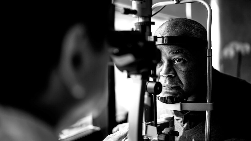
Optometrists are primary eye health professionals. Their job involves examining and managing problems with the eyes. Therefore, they play a major role in the early detection of eye conditions that can negatively affect vision, such as AMD.
An optometrist can check for eye disease and will typically do so through a
If an optometrist suspects AMD, they will suggest further tests to confirm a diagnosis and determine how advanced the damage is. Some optometrists may be able to perform these tests in their office, while others may choose to refer a person to an ophthalmologist for further examination and treatment.
Learn more about other types of eye care practitioners.
To maintain proper eye health, it is advisable for people to attend regular eye exams with an optometrist. This is particularly true for older adults, as conditions such as AMD become more common with age. Health experts advise that people ages 65 years and older should receive eye exams every year to support proper eye health.
In some cases, an optometrist may be able to recommend how often a person should undergo an eye exam.
If an optometrist suspects that a person may have AMD based on the results of a dilated eye exam, they will recommend further testing. Typically, this will involve the following tests:
Optical coherence tomography (OCT)
OCT is a noninvasive imaging scan that uses light waves to take detailed images of the eye. Eye care practitioners can use high resolution cross-sectional images of the inside of a person’s eye to diagnose a number of medical conditions, including AMD.
To prepare a person for an OCT scan, an eye doctor may use dilating eye drops to widen the person’s pupils. The person will sit in front of the OCT machine, rest their head on a support, and stay still. Over the next 5–10 minutes, the equipment will scan the person’s eyes to create the image.
Learn more about OCT for AMD.
Fluorescein angiography
Fluorescein angiography is another diagnostic imaging technique that involves using a special dye and camera to view the back of the eye.
This test usually takes less than 30 minutes and involves using dilating drops to widen the pupils. A nurse or technician will then inject a yellow dye, known as fluorescein, into a vein in the person’s arm. The dye will eventually reach the blood vessels in the eye.
Fluorescein can glow brightly after exposure to specific wavelengths of light. When the dye passes through the retina in the eye, a special camera will take pictures that will allow the eye doctor to observe any problems.
Other tests
Other tests that an eye care specialist may use when diagnosing AMD include:
- Optical coherence tomography angiography: This quick test combines OCT and fluorescin angiography to take pictures of the blood vessels in the retina. It does not involve the use of a dye.
- Indocyanine green angiography (ICGA): This is an imaging technique similar to fluorescin angiography that also uses a dye. This method can help identify drusen, which can be a risk factor for macular degeneration.
- Amsler grid: This is a simple exam that involves a square grid with a dot in the middle. If a person notices any distortion of the grid, such as blurry lines, wavy lines, patches of darkness, or patches of missing squares, this may suggest vision problems. However, an Amsler grid typically helps monitor vision and is not particularly helpful for diagnosis.
Learn more about AMD diagnosis.
In some cases, an eye care specialist may also recommend genetic testing. This will typically involve taking and testing a blood sample.
While anyone can develop AMD, certain genetic factors can significantly influence when the symptoms may start and how AMD might progress.
This is not currently common practice, as no gene therapy treatments are presently available. But as research and investigations continue, specific treatments may become available to successfully prevent or manage AMD.
The symptoms of AMD will depend on the stage of the disease. There are two types of AMD: dry and wet.
Dry AMD is the type that occurs first, and it happens in three stages: early, intermediate, and late. It occurs when the macula becomes thinner with age, and it usually progresses slowly over several years.
Wet AMD is a less common and more severe type of late AMD. Any stage of dry AMD can progress to wet AMD, but health experts always refer to wet AMD as late stage. It occurs when abnormal blood vessels begin to grow in the back of the eye and damage the macula.
Early dry AMD does not cause any symptoms. Some people with intermediate dry AMD may not notice symptoms, either. However, others may begin to notice mild symptoms, such as mild blurriness in their central vision or trouble seeing in low light.
Late AMD — either wet or dry type — may cause the
- blurry areas in the center of the vision
- blank spots in the vision
- difficulty seeing in low light
- straight lines appearing wavy
- colors seeming less vibrant
AMD occurs when the macula begins to thin. The macula is a small portion of the retina that is inside the back layer of the eye. It is responsible for central vision, color vision, and fine detail.
Health experts are currently unsure of the exact cause of AMD. However, they note that several factors relate to its development, including:
- older age
- genetics
- smoking
- inadequate nutrition
- high blood pressure
Treatment for AMD will
Currently, there are no treatment options for early AMD. An eye doctor may advise regular eye exams and certain lifestyle strategies, such as maintaining a nutritious diet, getting regular exercise, and quitting smoking, if applicable.
Read on to learn more about tips for preventing or slowing the progression of AMD.
For intermediate dry AMD, a doctor may suggest certain dietary supplements that may help stop or slow the progression of AMD. These supplements will typically include:
Learn more about natural remedies for AMD.
There are currently no treatments available for late dry AMD. The only form of dry AMD that is treatable is dry AMD with geographic atrophy, which involves an area of cell loss in the retina.
For wet AMD, treatment options can include anti-VEGF medication and laser surgery. Anti-VEGF medications are injections that doctors can administer into the eyes to help reduce the number of abnormal blood vessels in the eyes. Laser surgery, which involves shining laser light into the eyes, may help seal leaky blood vessels.
Learn more about treatments for wet AMD.
An optometrist can play a role in diagnosing macular degeneration. Typically, an optometrist can detect AMD during a routine eye exam. They may be able to perform further tests to confirm a diagnosis, or they might refer a person to an ophthalmologist.
Early detection and management of AMD are important to help slow and possibly prevent progression of the disease. Because AMD is more common in older age, it is advisable for people ages 65 years and older to have an eye exam every year or two.