
Rhegmatogenous retinal detachment (RRD) is when the gel-like fluid inside the eye gets behind the retina and pushes it away from its supporting blood vessels.
RRD is one type of retinal detachment (RD). The retina is a layer of light-sensitive tissue at the back of the eye. RD occurs when the retina pulls away from the blood vessels that supply it with oxygen and nutrients.
As with all forms of RD, RRD is a medical emergency that requires immediate treatment to prevent vision loss or blindness.
This article provides an overview of RRD, including its causes, the outlook for those with the condition, and more.
According to the
- rhegmatogenous retinal detachment (RRD)
- tractional RD
- exudative RD
Rhegmatogenous retinal detachment is the most common type of RD.
In RRD, a small tear in the retina allows the gel-like fluid, or “vitreous,” inside the eye to get behind the retina. There, it pushes the retina away from its supporting blood vessels.
According to the
The American Academy of Ophthalmology (AAO) outlines some additional risk factors for RRD. These include:
- trauma to the eye or head
- previous eye surgeries,
including cataract surgery - extreme nearsightedness (myopia)
- previous viral infection of the retina (viral retinitis)
- retinal lesions
- underlying hereditary vitreoretinopathy, which is a genetic condition affecting the vitreous and retina
As the
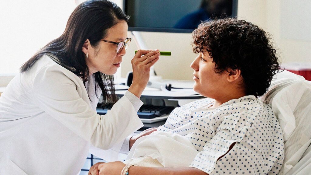
Anyone who experiences symptoms of RD should visit an ophthalmologist or an emergency room immediately.
Symptoms of RD often come on suddenly and may
- poor visual acuity, though some people may maintain excellent visual acuity
- flashes of light in one or both eyes
- a lot of new floaters, which are small dark spots or squiggles that float across the visual field
- a dark shadow or “curtain” that obscures part of the visual field
RRD is a medical emergency. Individuals with RRD symptoms should visit an eye doctor, or ophthalmologist, the same day their symptoms develop.
Surgical treatment for RRD aims to reattach the retina and restore vision. This may include one or more of the following procedures:
- Barrier laser retinopexy: This procedure is for localized, less severe detachments. Experts use a laser to create small burns around the retinal tear. This causes scarring that acts like a barrier and helps prevent the retina from detaching further. The patient is usually awake with topical anesthesia.
- Pneumatic retinopexy: An eye doctor injects an expanding gas bubble into the eye. The gas bubble floats over the detached area and pushes it against the back of the eye. The doctor then uses a freezing device to seal the retina against the wall of the eye.
- Scleral buckling: This procedure involves pushing the retina back into its supporting tissues so it has a chance to reattach.
- Vitrectomy: This procedure involves removing the vitreous and replacing it with saline or another solution.
Below are some answers to frequently asked questions about RRD.
Is rhegmatogenous retinal detachment serious?
As the
How do you diagnose rhegmatogenous retinal detachment?
According to a
As the AAO explains, OCT is a noninvasive imaging test that uses light waves to take cross-sectional images of the retina. The test allows ophthalmologists to view and accurately measure each layer of the retina.
What is the outlook for rhegmatogenous retinal detachment?
RRD is a medical emergency. Individuals with RRD symptoms should visit a healthcare professional the same day that their symptoms develop.
Treatment for RRD involves surgery. The surgery aims to reattach the retina and restore vision.
According to a
Rhegmatogenous retinal detachment (RRD) is a type of retinal detachment (RD) in which the vitreous of the eye gets behind the retina and pushes it away from its supportive blood vessels.
The condition may cause symptoms such as flashes, floaters, or dark shadows in the visual field.
As with other types of RD, RRD is a medical emergency that requires immediate treatment to prevent vision loss or blindness.
As such, anyone who experiences symptoms of RRD should visit an ophthalmologist or emergency room immediately for a diagnosis and appropriate treatment.
The treatment of RRD involves surgery to reattach the retina. Around 10% of such surgeries result in recurrent RDs that require additional interventions. Most of these recurrent RDs occur within 6 months of the initial surgery.