
Retrosigmoid craniotomy is a surgical approach that can treat brain tumors, lesions, and other health conditions that affect the posterior fossa, an area at the back of the skull.
Approaches like this one enable surgeons to access specific areas of the skull while allowing for fewer risks and shorter recovery times than open brain surgery. Surgeons may consider performing a retrosigmoid craniotomy to treat certain conditions, such as tumors, vascular malformations, and trigeminal neuralgia.
This article further discusses why a surgeon may perform a retrosigmoid craniotomy and what a person can expect before, during, and after the procedure.
A retrosigmoid craniotomy is a surgical approach that involves removing a small area of the skull, just behind the ear on either side of the head. The surgeon makes an incision behind a major blood vessel in the brain called the sigmoid sinus.
Surgeons use this technique to access the posterior fossa — the area at the back of the skull — and, more specifically, the cerebellopontine angle (CPA). The CPA is located between the cerebellum and the pons and is a common location for many posterior fossa tumors.
The retrosigmoid approach provides a clear view of and access to the following brain structures:
- the cerebellum
- the brain stem
- the facial nerve (cranial nerve VII)
- the vestibulocochlear nerve (cranial nerve VIII)
- certain blood vessels
A surgeon may need to access any of these structures depending on the reason for the procedure.
Learn more about the parts of the brain.
The retrosigmoid approach is a good alternative to more invasive brain surgeries. It requires removing a smaller part of the skull but still provides
There are several reasons why a surgeon may choose to perform this procedure, including:
- Tumor resection: Surgeons can use it to remove tumors or lesions in the CPA. The
most common of these are vestibular schwannoma, meningioma, and epidermoid cysts, which can cause symptoms such as headaches and hearing loss. - Trigeminal neuralgia: This is a condition that causes severe facial nerve pain. The
most common surgical treatment for trigeminal neuralgia is microvascular decompression, which aims to alleviate pressure on the trigeminal nerve. - Vascular malformations: This refers to an atypical structure of blood vessels that can occur in the brain as well as other parts of the body. Vascular malformations can lead to blood vessels becoming tangled or developing atypically, causing symptoms such as headaches, seizures, and bleeding.
- Facial nerve decompression: Compression of the facial nerve can occur for reasons such as injury or a tumor pressing on the nerve. It can cause symptoms such as weakened facial muscles, facial drooping, or paralysis. A surgeon may perform a retrosigmoid craniotomy to remove or correct structures pressing on the facial nerve.
Before having a retrosigmoid craniotomy, a person’s doctor will explain the procedure to them, discussing their specific surgical plan and what the individual can expect.
Before the procedure
The healthcare team will take a medical history and run certain tests to ensure the person is well-prepared for the surgery.
These tests may include:
- a neurological exam
- MRI scans
- CT scans
- PET scans
- angiography
- an electrocardiogram
- blood and urine tests
Before their surgery, a person will have multiple appointments in which they can discuss any medications they are taking with their surgical team and ask any questions.
In most cases, people will not be allowed to eat or drink anything on the day of their procedure. They will also need to follow any additional instructions their healthcare team gives them.
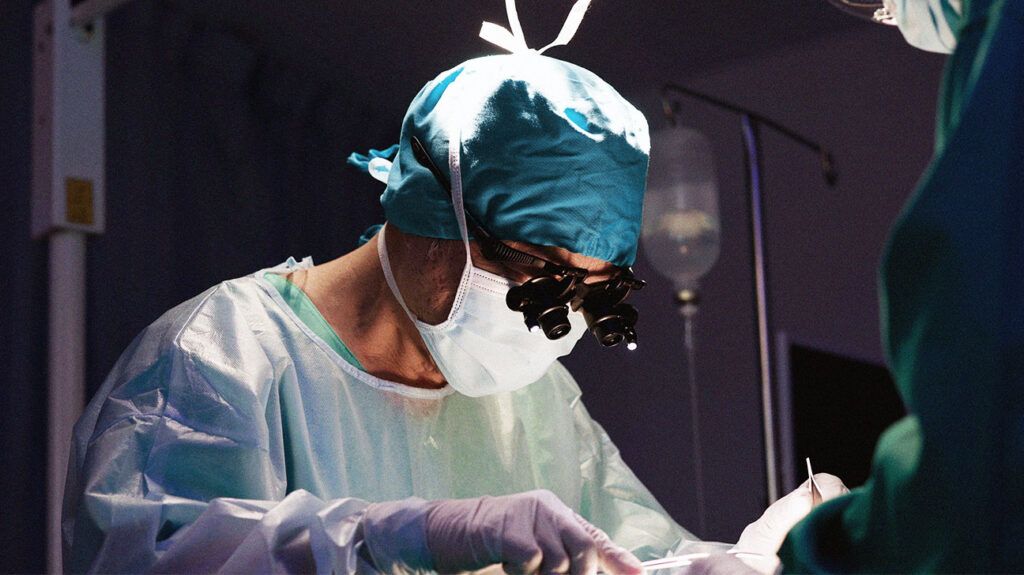
During the procedure
In some cases that require the person to be awake, the surgeon will administer a local anesthetic instead. Either way, the person should not feel any pain.
The surgical team may need to shave a small section of hair to access the surgical site. Then, the surgeon will make a small incision in a part of the head behind the ear and remove a small section of the skull to access the brain.
After the procedure, the surgeon will replace this part of the skull.
After the procedure
People usually wake up soon after the procedure is complete, although in some cases, a surgeon will keep the person asleep for longer to allow recovery.
It is normal to feel some soreness or discomfort after a craniotomy. Feeling tired after waking up from general anesthesia is common, too. A person’s healthcare team can provide pain relief, monitor their status, and help them stay comfortable after surgery.
The healthcare team may need to complete certain tests after surgery to assess the outcome and evaluate if it is necessary to organize physical, occupational, or speech and language therapy.
A person will usually stay in the hospital for up to 10 days after a craniotomy so that they can rest and recover with the help of their healthcare team. Generally, the full recovery period is around 6–12 weeks.
While a person is recovering, they will not be able to perform certain activities, such as:
- driving
- participating in contact sports or strenuous physical activity
- traveling by air
People will attend follow-up appointments after their surgery, where they can ask for advice from their healthcare team about what activities are safe for them to participate in.
Generally, the outlook for someone after a craniotomy will depend on many factors, such as the reason for their procedure and how effective the surgery was.
Many people fully recover and go on to resume their typical activities after having a craniotomy.
A
Major complications are not common following a craniotomy, occurring
Possible complications and side effects of a retrosigmoid craniotomy include:
- infection
- swelling
- scarring or denting at the surgical site
- vasospasm or artery constriction, which can narrow blood vessels
- a leak of cerebrospinal fluid, which surrounds the brain and spinal cord
- bleeding
- brain injury
- brain swelling
- brain hemorrhage
- neurological deficits or issues with the functioning of a certain body part
- speech difficulties
- damage to facial nerves
- damage to sinuses
- memory problems
- blood clots
- hearing loss
- balance and coordination problems
- headaches or migraine
- stroke
- coma
According to a 2020 study, surgery in the posterior fossa part of the brain can cause complications up to
Retrosigmoid craniotomy refers to a surgical approach that a surgeon uses to access a part of the brain at the back of the skull called the cerebellopontine angle. They may use this method for tumor resection or to treat other conditions.
A retrosigmoid craniotomy is a major neurosurgical procedure and requires a period of recovery during which a person may experience some side effects or complications. However, complications tend to be minor, and many people can resume their typical life activities after this procedure.